Dental Health Screening
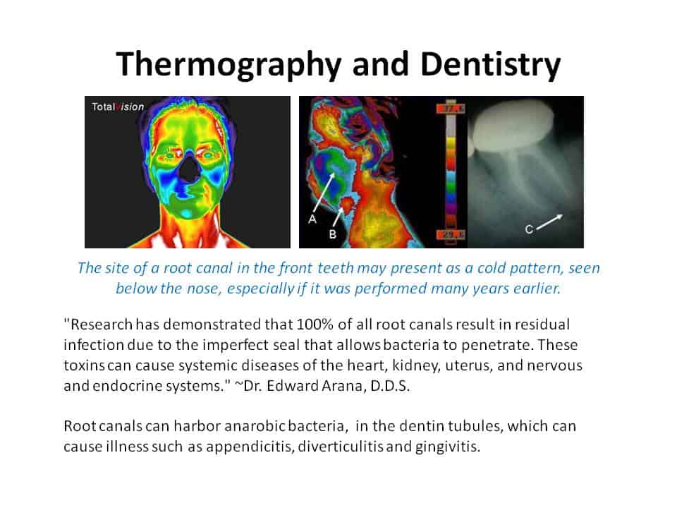
Thermal imaging, also called thermography and is not new in dentistry. The first articles about thermography in dentistry appeared in the 1970s. A thermography camera picks up infrared heat emissions and displays the result in a colored picture where each color represents a certain temperature.

Thermal imaging is an effective, radiation-free way to examine oral health issues and pain conditions. A healthy jaw will have even heat patterns throughout, while inflammation or infection will appear in higher temperatures on a thermal image. Dental thermography can more accurately identify facial pain, reducing unnecessary root canal treatments or extractions and other invasive treatments.
TMJ syndrome, implants, facial nerve injuries, and facial pain can all be further assessed and monitored through the use of dental thermography without the risks inherent in dental X-rays. Thermal images can even be used to monitor the progress of treatment.
If you have Temporal- mandibular Joint Disorder (TMD or TMJ), your jaw muscles are chronically strained. That is the reason you have TMD symptoms like facial pain and headaches. The jaw muscles take up a lot of the area on each side of the head and stress causes them to become inflamed.
Normally, the body is symmetrical in the amount of infrared radiation it gives off. If you have TMD on one side only, that will show up clearly in your thermal images. If you have it in both jaw joints, that will also be clear, each side showing its unique pattern of stress. Thermal imaging is highly sensitive to any abnormality in the muscles, blood vessels, nerves, and bones.
Dentists recommend the use of Medical Thermography in monitoring control in the inflammation process into oral cavity and reaction of the regional lymphatic nodes, maxillary joint disease and other chronic diseases of the bones, nerves, located in the maxilla facial area. Medical Thermography can also measure temperature changes in the application of new methods and dental materials applied by dentists.
HOW IS THERMOGRAPHY USED?
Different types of body tissue have slightly different temperatures. When tissue is abnormal or inflamed, a thermography camera can record its temperature as a color distinct from the color of surrounding healthy tissue. Inflammation creates heat. If part of the body is inflamed, it will look different in the thermographic image (thermogram).
Disclaimer: Carolina Medical Thermography is not a treatment or diagnosing center. The information at this website is for general information and resource purposes only and is not intended in any way to be a substitute for professional medical advice, diagnosis or treatment.

